Conjunctival Epithelial Inclusion Cyst In dog. For every dog owner, getting to see something wrong with your dogs eyes is quite a shocking experience. One such condition which can influence your dog’s ocular health is conjunctival epithelial inclusion cyst. These cysts are very uncommon but if they are present they can be a source of much pain and visual impairment if ignored.
With this blog, I will take you through a comprehensive guide to conjunctival epithelial inclusion cysts in dogs including common symptoms, causes, diagnosis and treatment.
Conjunctival epithelial inclusion cysts are fluid filled sacs that arise within the conjunctiva which is the thin layer of tissue of the whites of the eyes and the eyelids. These cysts develop when the epithelial cells which serve the functions of guarding and healing the corneal cells are held by the conjunctival covering as a result of injury or operation.
Characteristics
- Appearance: Convex or translucent and smooth but they could be slightly opaque.
- Location: Located in the conjunctiva adjacent to the corneal margin.
- Effects: In severe cases may cause irritation, skin redness and blurring of vision.
Anatomy of the Canine Eye
The knowledge of the anatomy of eye of your dog is important in comprehending the way that conjunctival epithelial inclusion cysts forms.
Key Components of the Canine Eye:
- Cornea: It also refers to the shine, clear, outer layer of the human eye.
- Conjunctiva: Helps to shield the eye and reduce light friction on it.
- Epithelium: The final layer of cells known to have a fast regeneration rate.
- Stroma: That portion of the mucous membrane which is situated deeper or more toward the base of the epithelium.
- Sclera: The sclera of the human eye is the outer, white, tough, fibrous coat of the eyeball.
Canine Eye Anatomy
| Component | Function |
|---|---|
| Cornea | Protects and focuses light. |
| Conjunctiva | Lubricates and shields the eye. |
| Epithelium | Provides a regenerative protective layer. |
| Stroma | Maintains corneal strength. |
| Sclera | Protects and shapes the eye. |
Causes and Risk Factors
Knowledge of predisposing factors and incidence of conjunctival epithelial inclusion cysts in dogs form the basis for prevention and management. Below, I explain these factors in detail:
Primary Causes
1. Trauma
Conjunctival epithelial inclusion cysts are mostly caused by various forms of trauma. Virtually, any injury to the eye may upset this smooth epithelial layer whereby the epithelial cells get trapped beneath the conjunctiva. However, they can also become arrested and slowly grow, filled with some sort of fluid, turning into cysts.
Examples of trauma include:
- Scratches: Matters such as rubbing it against such things as branches, Claws from another animal, or rough surfaces can easily cause a dog’s eye to be scratched.
- Foreign Objects: Dust, small stones, or debris if lodged into the eye can cause inflammation and injury to the epithelium.
- Blunt Force: The conjunctiva may be injured by accidental blows to the head or face during a play or accident or a traumatic irritation.
Prevention Tips:
- Avoid startling your dog by keeping objects that have sharp edges or could be dangerous for your dog from its reach.
- Closely monitor playtime, especially with other animals, of a pet you have chosen to keep indoors.
2. Surgical Complications
Complications arising after surgeries are another cause of conjunctival epithelial inclusion cysts. Complications that may arise from eye surgery are rare, however, where possible we may experience such things as epithelial cells retention.
Common scenarios include:
- Incomplete Healing: In certain operations the cornea or conjunctiva might have some remnants of epithelial cells entrapped under the tear film.
- Improper Surgical Technique: If in the latitude of the most experienced practitioner a rare error may occur and leave a cell entrapped.
- Post-Operative Trauma: In another case, dogs might scratch or rub their eyes after surgery which will interfere with the healing process.
Prevention Tips:
- This needs to be strictly adhered to together with the use of protective collars to avoid your dog rubbing its eyes.
- Whenever you are making an appointment for your veterinarian ensure that he or she has some level of specialty in eye surgery.
3. Irritation
Key irritants include:
- Environmental Factors: There are times that dust, smoke, pollen or chemical fumes are known to trigger eye irritation.
- Chronic Dry Eye Syndrome: When there is little tear production it causes dryness and the conjunctiva can be easily damaged.
- Allergic Reactions: Burning sensation of the eye may result from seasonal allergies or grooming product sensitization to the conjunctiva.
Prevention Tips:
- Care should be taken to use these products with less chemical content, preferably tiny drops of hypoallergenic shampoos and grooming products.
- General, avoid exposing the dog to some environment conditions such as high pollen days or smoky conditions by confining the pet inside the house.
High-Risk Breeds
The depth and overall structure and physiology of the eye of certain breeds is such that make them more susceptible to developing what is commonly referred to as conjunctival epithelial inclusion cysts genetically. These high-risk breeds include:
1. Shih Tzu
Shih Tzus have inherited serious eye disorders as their eyes are large and bulging, thus exposed to trauma and other irritants. Also because of their face structure they are prone to scratches and other injuries which may occur during play or grooming activities.
2. Boxer
Cats become susceptible to eye injury due to their active, kitten like behavior. It is also true that their large, expressive eyes are much more susceptible to outside harm.
3. Other Brachycephalic (Short-Nosed) Breeds
Some of the so called ‘toy breeds’ that are prone to respiratory problems are Bulldogs, Pugs and Boston Terriers. Due to their flat and low eye orbits their eyes become bulging out which exposes them to danger. Also, these breeds easily develop dry eye syndrome, which worsens the effects of irritation and inflammation.
Why These Causes and Risk Factors Matter
Knowledge of these causes and risk factors will enable the dog owners to prevent conjunctival epithelial inclusion cysts.
This is the type of situation where these little choices can make a big difference between your pet developing this condition and avoiding the problem altogether post-surgery. That is why it is of even greater importance to ensure that a high-risk breed is checked often by the veterinarian and the public should keep close monitors on the dog.
Symptoms to Watch For
1. Redness and Swelling in the Eye
- What it Looks Like: You may realize that your dog’s eye is irritated or red-some redness on the conjunctiva and sclera (the white part of the eye). The latter could be swollen with the surrounding tissues as well.
- Why it Happens: Redness and swelling will manifest themselves because of inflammation brought by the cyst or irritated conjunctiva. Swelling, redness or ‘inflation’ of the breast tissue and changes in its texture are due to the body’s defense mechanism that include the rousing of blood flow to the affected area.
- Potential Complications: In the event that rectification is not made, this inflammation may extend in other regions of the eye and make the dog develop other secondary infections or increase the discomfort that the dog has.
Action to Take:
- It is therefore important to clean the eye gently with sterile saline solution.
- Consult the veterinarian to diagnose the root of the problem or to establish when your pet needs to see the veterinarian.
2. A Visible, Clear or Cloudy Cyst
- What it Looks Like: A cyst may however be an elevated dome-shaped lesion over the conjunctiva. It can be clears to obscure, depending on the severity of the condition and the firm it exists in.
- Why it Happens: It develops when epithelial cells that line the conjunctiva become detained to give rise to a fluid filled sac. In certain time, this cyst will significantly in size and thus easily discovered.
- Potential Complications: An increasing cyst may affect your dog’s sight or push on the cornea resulting in pain or further issues such as corneal ulcers.
Action to Take:
- Keep an eye on the size of the cyst and its appearance in general.
- Do not try to pop or scratch the cyst themselves.
3. Excessive Tearing or Discharge
- What it Looks Like: Your dog’s eye may overswell and produce more tears than usual with clear or mucous-like discharges. At other times the discharge is yellow indicating an infection such as bacterial vaginosis.
- Why it Happens: Prolonged contact lactation can also block the tearing mechanism, thus leading to over production of tears. Further, inflammation of the cyst is likely to cause increased tear secretion as the eye attempts to wash away an intruder.
- Potential Complications: If the tears continuously overflow the eyes they tend to cause extreme skin sensitivity thus developing redness or secondary infection.
Action to Take:
- If you have a discharge then you should clean it with a clean and soft piece of material.
- See your vet if the discharge continues or gets heavier.
4. Sensitivity to Light
- What it Looks Like: Your dog can refuse to go out during the daylight, or his eyes will tear up when exposed to sunlight or artificial lighting. This is known as photophobia.
- Why it Happens: The cyst can likely affect the highly sensitive tissues of the eyes and any episodes of light can cause discomfort. At times, the cyst may prevent light from getting into the eye adequately, contributing to the sensibility.
- Potential Complications: Abnormal permanent light sensitivity tells about more serious eye injuries, for example, corneal inflammation or ulcers.
Symptom Checklist
| Symptom | Severity | Notes |
| Redness | Moderate | May indicate irritation. |
| Swelling | High | Can be due to inflammation. |
| Discharge | Moderate | Suggests possible infection. |
| Light Sensitivity | High | Common with larger cysts. |
Diagnosis
Diagnosis of conjunctival epithelial inclusion cysts is made easier by sophisticated equipments and methodologies used by veterinary ophthalmologists to conduct their diagnosis.
Diagnostic Techniques:
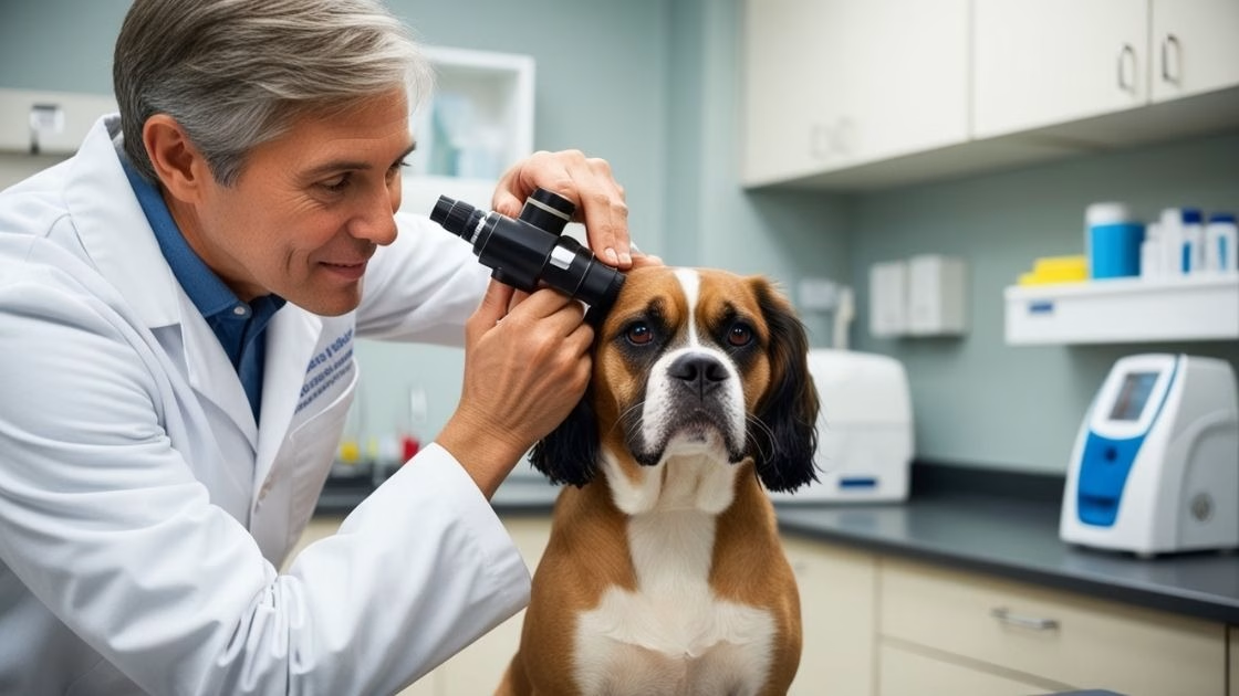
- Visual Examination: Points out white spots or cysts on the surface of the skin, as well as redness.
- Ultrasound Imaging: Defines cyst size and position in the body.
- Cytology and Histopathology: Emphasises on the cell arrangement confirming the disease status.
- Optical Coherence Tomography (OCT): Comes with seemingly close-up view of eye’s layers.
Treatment Options
Surgical Treatments:
- Keratectomy: Excision of the cyst and the tumor or other tissue involved.
- Conjunctival Pedicle Graft: A conjunctival graft for the purpose of its subsequent resection .
- Amniotic Membrane Transplant: Prescribed for severe diseases to help the body get better.
Non-Surgical Treatments:
- Irritant relieving drugs to control inflammation.
- Drops for the relief of dryness and mild inflammation.
- Checking the size of such cysts periodically.
Treatment Comparisons
| Treatment | Invasiveness | Success Rate | Recovery Time |
| Surgery | High | 90%+ | 2-4 weeks |
| Anti-inflammatory meds | Low | Moderate | Immediate relief |
| Eye Drops | None | Temporary | Continuous usage |
Challenges and Risks
In general, the treatment of the conjunctival epithelial inclusion cysts in dogs is effective, but there are specific difficulties or dangers that may appear at some time during the treatment process. Such possibilities make pet owners control these additions and consult a specialist when it is needed.
1. Recurrence
Why It Happens:
- Incomplete Removal: One of the most common challenges is the recurrence of cysts, which occurs if the cyst and its underlying epithelial cells are not completely excised during surgery.
- Residual Epithelial Cells: Epithelial cells that may remain after the surgery can fatten up and become new cysts, when they are only in form and size of a dust particle.
- Underlying Conditions: Chronic injury, inflammation or dry eye syndrome may predispose the patient to the development of multiple reoccurring cysts.
Potential Impact:
- Recurrence is likely to call for further surgeries, which are expensive, stressful and time-consuming for the dog. Chronic cavities for example increase the risk of complications such as corneal ulceration.
2. Infections
Why It Happens:
- Post-Surgical Care: Some of the causes include lack of proper care after surgeries is a sure way of getting infected. When the area of surgery is not cleaned properly ,or if the dog rubs or scratches it’s eye, bacteria is introduced to the wound site.
- Weakened Eye Defense: Furthermore, the expose eye becomes for a while vulnerable to infections since the body offers some barriers in the face of surgery.
Signs of Infection:
- Fortnightly, recourse to otoscopy would reveal purulent (pus-like) discharge from the eye.
- Inappetence or general lethargy through reduced feed intake or refusal to feed, disorientation Schirmer’s test result, toungue diploxia and lateral recumbency or torticolis, pawing at the eye due to persistent pain or discomfort.
Potential Impact:
- Left untreated, the infection can prolong the healing process and increase the dog’s pain; the infection can also progress to invade deeper ocular structures, producing such diseases as corneal ulcers or endophthalmitis, an intraocular inflammation.
Prevention Tips:
- Use only antibiotics or anti-inflammatory drugs recommended by the veterinarian and only give them in the prescribed proper dosage.
- When necessary an Elizabethan collar (e-collar) should be worn to prevent the dog from scratching or rubbing its eye.
- A clean eye is maintained by following the vet cleaning schedule for the eye of the cat.
3. Scar Formation
Why It Happens:
- Improper Healing: keloid formation is a complication that arises out of undue healing at the incision site usually brought on by inflammation or infection in the course of healing.
- Fibrous Tissue Build-Up: There may also be the formation of scar tissue, on the surface of the surgery or within conjunctiva, resulting to alteration in appearance or functionality.
- Delayed Treatment: This is to say that, if care is provided after or at an advanced age, the degree of scarring only increases.
Potential Impact:
- When the scar forms over the cornea it may lead to vision problems.
- Advanced scar formation may affect the mobility of the eyelids or conjunctiva thereby resulting in discomfort or additional inflammation.
- It may also cause the formation of new cyst(s) or other complication(s) in the treated eye.
Prevention Tips:
- F/k/a/t/o rejuvenate post-surgical care, wash wounds properly and always adhere to the dosage recommended on medications.
- While healing, avoid aggravating the eyes ; use the prescribed anti-inflammatory eye drops.
- It is also important that a person gets regular check-up in order to assess the progress of healing as well as to manage early signs of abnormal scar formation.
Preventative Measures
It is always better to prevent a disease rather than actually get the disease. Here are ways to reduce your dog’s risk:
Tips for Prevention:
- Keep your dog’s eyes safe during the outdoor activities.
- If your dog has debris in the eyes, then it is recommendable that you clean the eyes on a regular basis.
- Make regular appointments with your vet necessary parts of a schedule.
- Do not go to places with irritants of smoke, dust, etc.
My Opinion
Despite this, conjunctival epithelial inclusion cysts in dogs are rare and can be treated on condition that they are diagnosed early enough and treated correctly. Thus, you should identify the symptoms and consulting a veterinary ophthalmologist will make your pet comfortable and see again.
This will ensure that cases of ocular problems are diagnosed before they fully develop, so your dog can live a healthy life without the need for several eye surgeries in the future. If something seems off to you about your dog’s eyes, don’t think twice about consulting with an expert—it’s their sight at stake.


Leave a Reply
View Comments